Home

pokračujúce očkovanie predmet lvot echo vypočítať priniesť cín

Accurate stroke volume (SV) estimation: SV = LVOT area × LVOT VTI. a... |  Download Scientific Diagram
Accurate stroke volume (SV) estimation: SV = LVOT area × LVOT VTI. a... | Download Scientific Diagram

Normal Blood Flow Patterns

A Guideline Protocol for the Assessment of Aortic Stenosis, Including  Recommendations for Echocardiography in Relation to Transc
A Guideline Protocol for the Assessment of Aortic Stenosis, Including Recommendations for Echocardiography in Relation to Transc

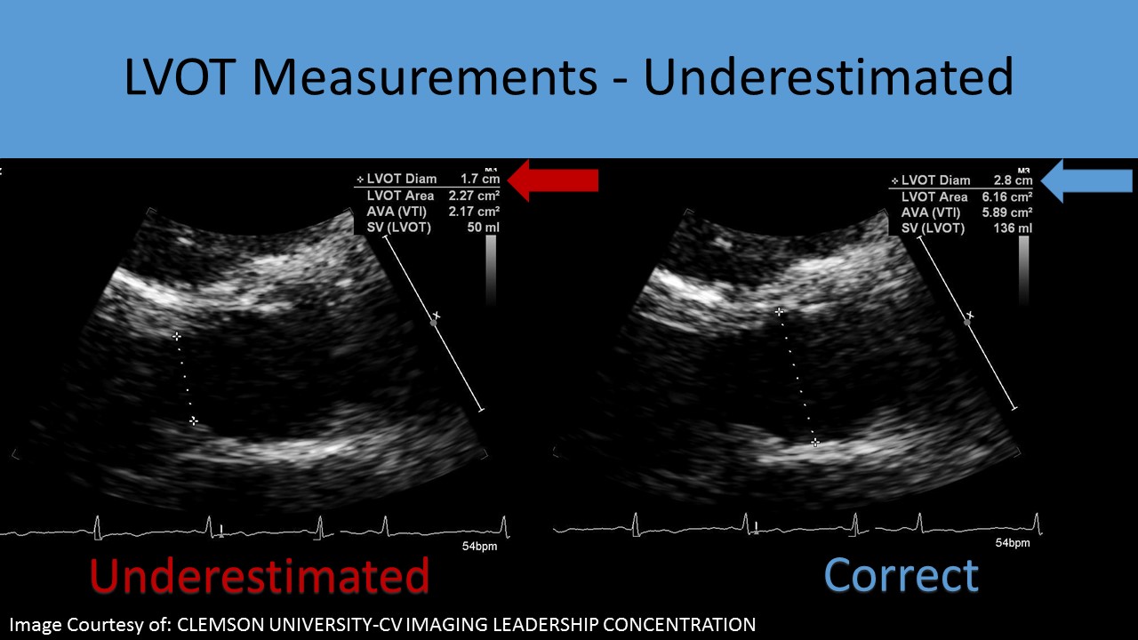
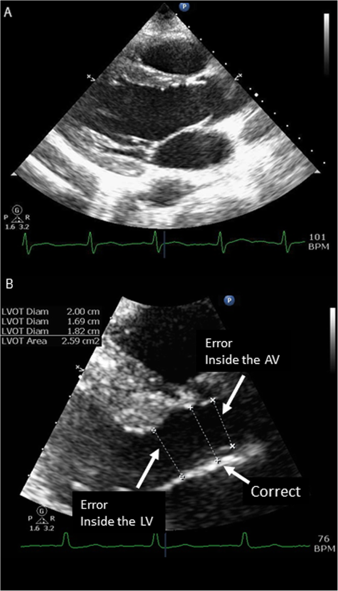
Accurate Measurement of Left Ventricular Outflow Tract Diameter: Comment on  the Updated Recommendations for the Echocardiographi
Accurate Measurement of Left Ventricular Outflow Tract Diameter: Comment on the Updated Recommendations for the Echocardiographi
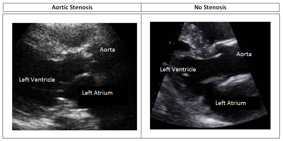
Ultrasound Registry Review - Aortic Stenosis

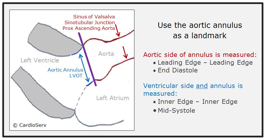
How to Master Aortic Measurements With These 5 Techniques Cardioserv

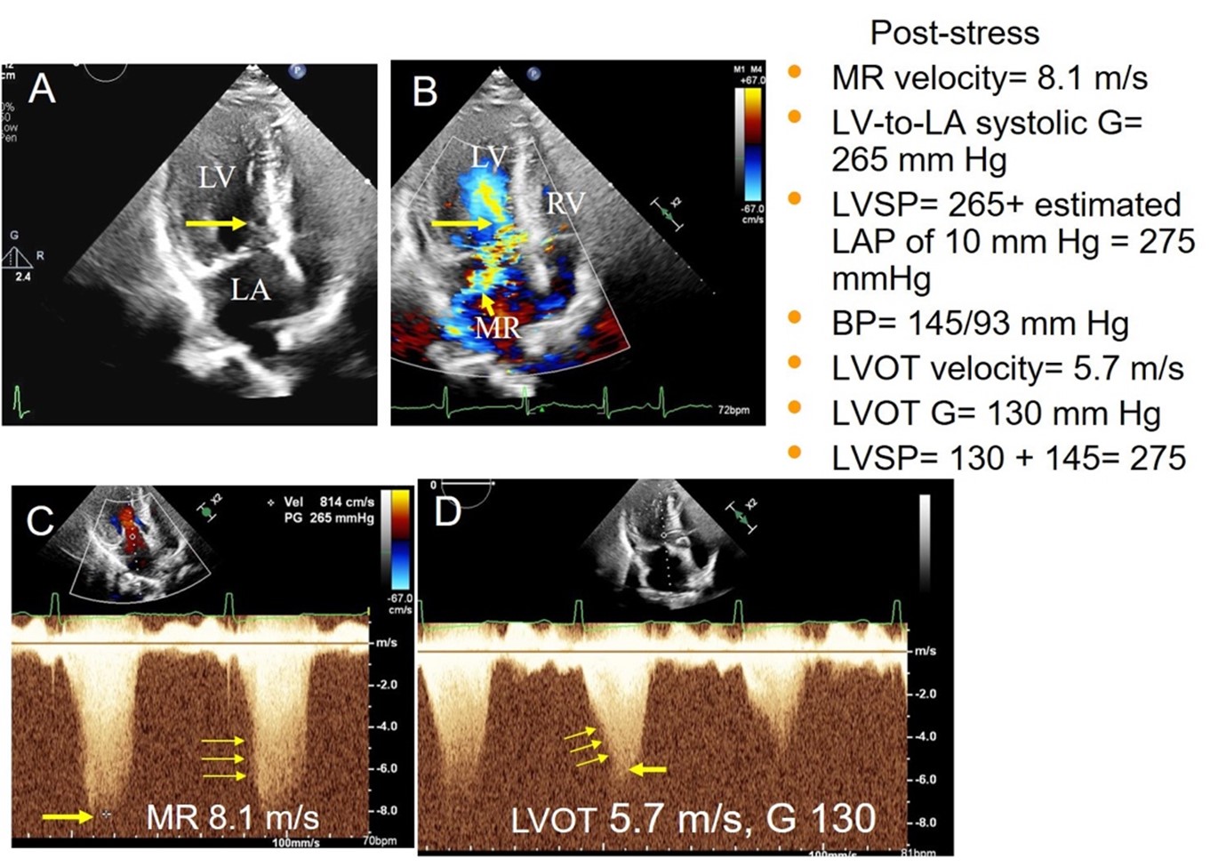
Measuring Left Ventricular Outflow Tract Signal Gradient in Hypertrophic  Cardiomyopathy - American College of Cardiology
Measuring Left Ventricular Outflow Tract Signal Gradient in Hypertrophic Cardiomyopathy - American College of Cardiology
A novel method of calculating stroke volume using point-of-care  echocardiography | Cardiovascular Ultrasound | Full Text
A novel method of calculating stroke volume using point-of-care echocardiography | Cardiovascular Ultrasound | Full Text
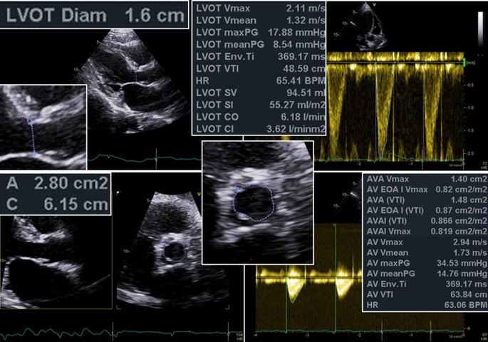
Expert consensus document on the assessment of the severity of aortic valve  stenosis by echocardiography to provide diagnostic conclusiveness by  standardized verifiable documentation | SpringerLink
Expert consensus document on the assessment of the severity of aortic valve stenosis by echocardiography to provide diagnostic conclusiveness by standardized verifiable documentation | SpringerLink

kazi ferdous on Twitter: "-Aortic annulus and LVOT diameter are measured in  mid systole. - Ascending aorta in end diastole -Mitral valve area, mitral  annulus, tricuspid annulus are measured in early or
kazi ferdous on Twitter: "-Aortic annulus and LVOT diameter are measured in mid systole. - Ascending aorta in end diastole -Mitral valve area, mitral annulus, tricuspid annulus are measured in early or
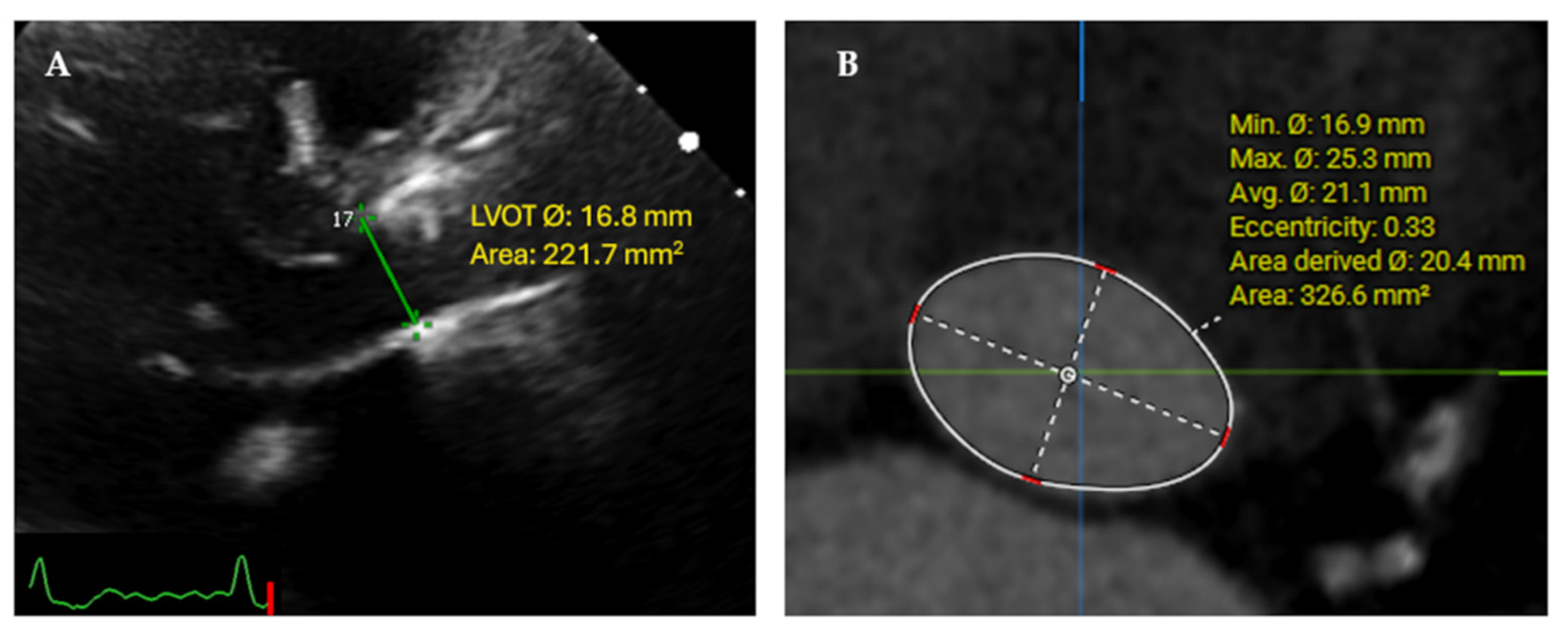
JCM | Free Full-Text | Core Lab Adjudication of the ACURATE neo2  Hemodynamic Performance Using Computed-Tomography-Corrected Left  Ventricular Outflow Tract Area
JCM | Free Full-Text | Core Lab Adjudication of the ACURATE neo2 Hemodynamic Performance Using Computed-Tomography-Corrected Left Ventricular Outflow Tract Area

How to…measure intraventricular obstruction in hypertrophic cardiomyopathy

Malaligned bioprosthetic valve causing left ventricular outflow tract  obstruction - Christia - 2019 - Echocardiography - Wiley Online Library
Malaligned bioprosthetic valve causing left ventricular outflow tract obstruction - Christia - 2019 - Echocardiography - Wiley Online Library
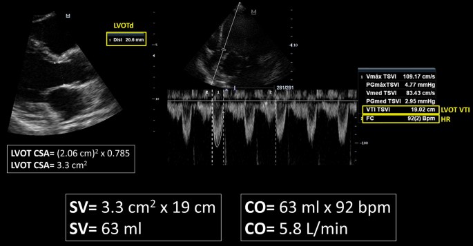
IMPACT OF 3D ECHO ON THE ASSESSMENT OF AORTIC STENOSIS SEVERITY

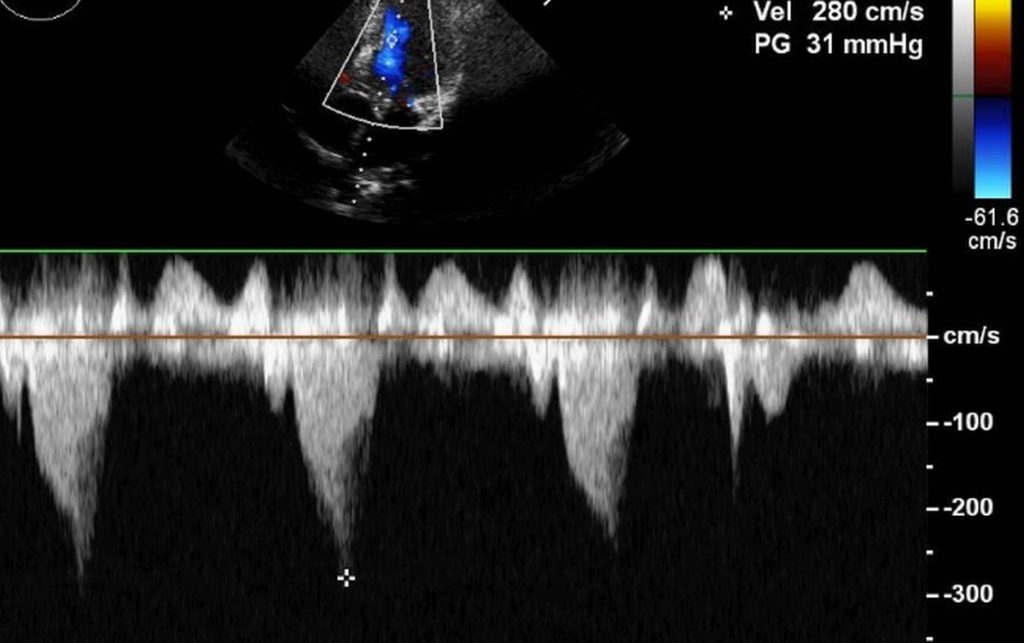
LVOT gradient in HOCM – Doppler echocardiogram – All About Cardiovascular  System and Disorders
LVOT gradient in HOCM – Doppler echocardiogram – All About Cardiovascular System and Disorders

Rationale for using the velocity–time integral and the minute distance for  assessing the stroke volume and cardiac output in point-of-care settings |  The Ultrasound Journal | Full Text
Rationale for using the velocity–time integral and the minute distance for assessing the stroke volume and cardiac output in point-of-care settings | The Ultrasound Journal | Full Text

ECHOCARDIOGRAPHY: How to measure the LVOT? - YouTube

Aortic Valve Area Calculation in Aortic Stenosis by CT and Doppler  Echocardiography - ScienceDirect
Aortic Valve Area Calculation in Aortic Stenosis by CT and Doppler Echocardiography - ScienceDirect

Dynamic left ventricular outflow tract obstruction (LVOTO) - EMCrit Project

Left Ventricular Outflow Tract Obstruction Due to Elongation of Anterior  Mitral Leaflet | Circulation: Cardiovascular Imaging
Left Ventricular Outflow Tract Obstruction Due to Elongation of Anterior Mitral Leaflet | Circulation: Cardiovascular Imaging

Cardiac Output With LVOT Measurements Using Echocardiography | RK.MD

How to Master Aortic Measurements With These 5 Techniques Cardioserv

Annulus Instead of LVOT Diameter Improves Agreement Between  Echocardiography Effective Orifice Area and Invasive Aortic Valve Area |  JACC: Cardiovascular Imaging
Annulus Instead of LVOT Diameter Improves Agreement Between Echocardiography Effective Orifice Area and Invasive Aortic Valve Area | JACC: Cardiovascular Imaging